
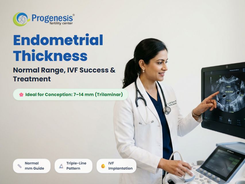
In human reproduction, successful conception requires two equally critical elements: a genetically healthy embryo (the "seed") and a receptive, nourished uterine lining (the "soil"). The medical term for this vital uterine foundation is the endometrium, and its depth—known clinically as endometrial thickness—is one of the most reliable predictors of natural pregnancy, embryo implantation, and In Vitro Fertilization (IVF) success.
Whether you are preparing for a natural conception cycle, Intrauterine Insemination (IUI), or a Frozen Embryo Transfer (FET), understanding your endometrial thickness report can help you navigate your fertility jouey with clarity and confidence.
In this comprehensive clinical guide, senior reproductive endocrinologists and fertility consultants at Progenesis Fertility Center break down what constitutes normal endometrial thickness across different menstrual cycle phases, the significance of the "trilaminar" triple-line pattern, what causes an abnormally thin or thick lining, and advanced evidence-based treatments to optimize your uterine environment for a healthy pregnancy.
What is Endometrial Thickness?
The endometrium is the innermost mucosal epithelial lining of the uterus. Throughout a woman’s reproductive years, this dynamic tissue undergoes continuous hormonal remodeling: it thickens in response to estrogen, matures under the influence of progesterone, and—in the absence of conception—breaks down and sheds during menstruation.
Endometrial thickness refers to the cross-sectional measurement of this inner lining, typically assessed in millimeters (mm) via high-resolution transvaginal ultrasound (TVS).
Clinical Implantation Fact: For successful embryo implantation, fertility specialists evaluate both quantity (thickness in millimeters) and quality (ultrasound texture and vascular perfusion). An ideal lining must be sufficiently thick and architecturally organized to welcome and sustain an implanting blastocyst.
Also Read: Everything You Need to Know About IVF Treatment & Success Rates
How is Endometrial Thickness Measured?
Ultrasound imaging is the gold-standard, non-invasive method for measuring uterine lining thickness:
- Transvaginal Ultrasound (TVS): A specialized ultrasound probe is placed in the vagina, providing high-frequency, crystal-clear imaging of the pelvic structures. The sonologist captures a longitudinal (sagittal) view of the uterus.
- Double-Layer Measurement: The caliper measures from the outer echogenic border of the anterior endometrial stripe across the uterine cavity to the posterior border at its thickest point (fundal region). This represents the full double-layer thickness.
- Pelvic MRI (Special Cases): When severe uterine retroversion, large uterine fibroids, or extensive adenomyosis distort ultrasound visualization, pelvic magnetic resonance imaging (MRI) provides precise cross-sectional measurements.
Endometrial Thickness Across Menstrual Cycle Phases
Your uterine lining is never static. Its depth varies predictably across the 4 biological phases of your menstrual cycle:
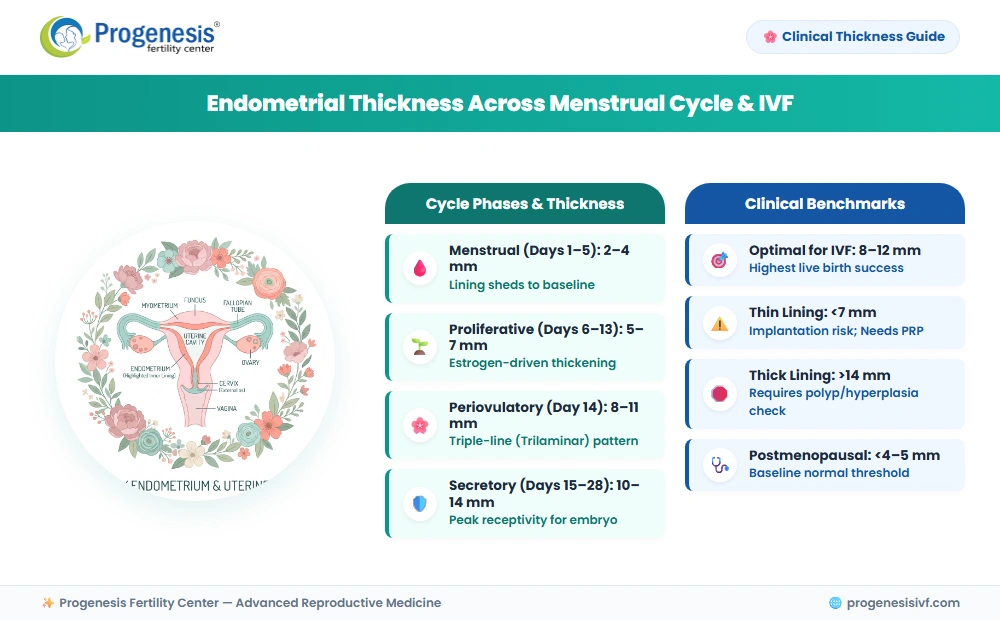
Phase-by-Phase Endometrial Thickness Breakdown:
| Menstrual Phase | Cycle Timing | Normal Thickness (mm) | Ultrasound Architecture | Key Hormonal Driver |
|---|---|---|---|---|
| Menstrual Phase | Days 1 to 5 | 2 to 4 mm | Thin, broken echogenic stripe as functional layer sheds with menstrual flow. | Low Estrogen & Progesterone baseline |
| Early Proliferative Phase | Days 6 to 10 | 5 to 7 mm | Smooth, thin, hypoechoic single layer as tissue regenerates. | Rising Estradiol (E2) from growing ovarian follicles |
| Late Proliferative (Periovulatory) | Days 11 to 14 | 8 to 11 mm | Classic "Trilaminar" (Triple-Line) pattern with distinct outer and central echogenic lines. | Peak pre-ovulatory Estrogen surge |
| Secretory (Luteal) Phase | Days 15 to 28 | 10 to 14 mm | Homogeneous, bright hyperechoic texture; glands swell with glycogen and nutrients. | Progesterone produced by the Corpus Luteum |
| Postmenopausal (Non-bleeding) | Baseline | < 4 to 5 mm | Atrophic, uniform, thin white line. | Low postmenopausal hormone baseline |
What is the Ideal Endometrial Thickness for Pregnancy & IVF?
One of the most frequent questions asked by fertility patients is: "What endometrial thickness is needed to get pregnant?"
Extensive reproductive clinical studies indicate that 8 to 12 mm is the sweet spot associated with the highest clinical pregnancy and live birth rates in both natural conception and assisted reproductive cycles.
Clinical Implantation Probability Matrix:
| Thickness Category | Measurement (mm) | Implantation Probability | Clinical Recommendation |
|---|---|---|---|
| Severely Thin | < 6.0 mm | Significantly Reduced (<15%) | Transfer cancellation strongly considered; evaluate for uterine scarring or vascular insufficiency. |
| Borderline / Suboptimal | 6.0 to 6.9 mm | Moderate (20% – 30%) | Acceptable if triple-line pattern is distinct, but individualized protocol optimization advised. |
| Optimal Window | 8.0 to 12.0 mm | Maximum Peak (55% – 70%) | Ideal for natural conception and embryo transfer; highest live birth rates. |
| Adequate / Safe | 12.1 to 14.0 mm | High (50% – 60%) | Favorable, provided fluid and intracavitary polyps are absent. |
| Excessively Thick | > 14.0 to 15.0 mm | Reduced / Variable | Evaluate for endometrial hyperplasia, polyps, or unopposed estrogen stimulation. |
The Critical Role of the "Trilaminar" (Triple-Line) Pattern
Thickness alone is not the sole determinant of uterine receptivity. Fertility specialists evaluate the endometrial echopattern:
- Trilaminar Pattern (Triple-Line): Appears just before ovulation or prior to starting progesterone in an IVF cycle. It consists of three parallel echogenic lines separated by two hypoechoic layers.
- Why It Matters: A 7.5 mm lining with a crisp triple-line appearance often produces higher pregnancy rates than a 9 mm lining that appears prematurely dense, hazy, or hyperechoic.
Also Read: What is Bulky Uterus? Symptoms, Causes, and Treatments
Thin vs Thick Endometrium: Symptoms, Causes & Clinical Fixes
When the endometrial lining fails to develop properly or grows uncontrollably, it can impair fertility and cause irregular vaginal bleeding.
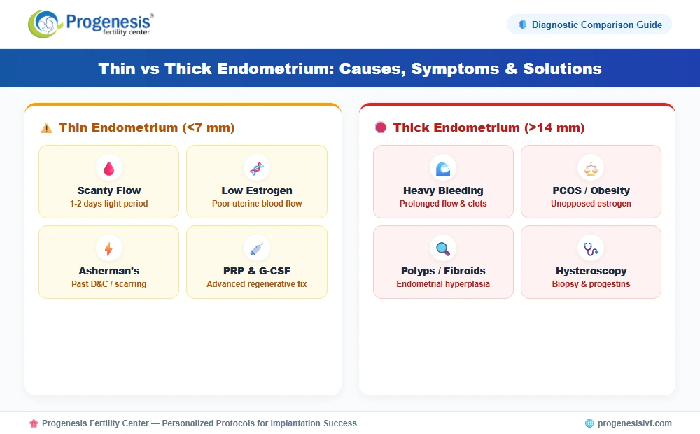
Comparison: Thin Endometrium vs Thick Endometrium
| Clinical Parameter | Thin Endometrium (< 7 mm) | Thick Endometrium (> 14 mm) |
|---|---|---|
| Primary Symptoms | Very light/scanty periods (hypomenorrhea), 1–2 days bleeding, difficulty conceiving, recurrent implantation failure. | Heavy menstrual bleeding (menorrhagia), prolonged periods (>7 days), passing large blood clots, bleeding between cycles. |
| Hormonal Profile | Suboptimal estrogen levels, poor ovarian response, or resistant estrogen receptors. | Estrogen dominance without sufficient progesterone counter-balance (common in PCOS and obesity). |
| Common Anatomical Causes | Asherman's syndrome (intrauterine adhesions from past D&C), chronic endometritis, pelvic tuberculosis. | Endometrial polyps, submucosal fibroids, endometrial hyperplasia, hormone replacement therapy without progestins. |
| Implantation Impact | Embryo cannot establish vascular connection; high risk of chemical pregnancy or miscarriage. | Irregular architecture disrupts window of implantation; risk of underlying cellular atypia. |
| Primary Clinical Solution | Escalated estrogen, vasodilators, intrauterine PRP infusions, G-CSF, hysteroscopic adhesiolysis. | Diagnostic hysteroscopy with endometrial biopsy, progestin therapy (oral or Mirena IUD), polyp removal. |
Proven Treatments to Increase Endometrial Thickness
If you have been diagnosed with a thin endometrium during an IUI or IVF cycle, do not lose hope. Advanced reproductive medicine offers targeted, evidence-based interventions to stimulate endometrial growth:
1. Hormone Optimization Protocols
- Extended Estrogen Administration: Utilizing multiple delivery routes (oral micronized estradiol, transdermal patches, or vaginal estradiol tablets) provides higher, sustained local tissue concentrations to stimulate cell proliferation.
- Low-Dose hCG Priming: Small doses of human chorionic gonadotrophin administered during the follicular phase can stimulate local angiogenic growth factors.
2. Enhancing Pelvic & Uterine Blood Flow
- Vasodilators (Sildenafil / L-Arginine): Vaginal sildenafil suppositories and oral L-arginine enhance nitric oxide pathways, dilating radial uterine arteries to improve endometrial perfusion.
- Low-Dose Aspirin & Vitamin E: Improves microcirculation and provides antioxidant protection to developing endometrial endothelial cells.
3. Advanced Regenerative Therapies
- Intrauterine Platelet-Rich Plasma (PRP) Infusion: Autologous blood is centrifuged to concentrate growth factors (VEGF, PDGF, TGF-beta) and infused into the uterine cavity 48–72 hours prior to embryo transfer. Clinical studies show significant improvement in refractory thin endometrium.
- Granulocyte Colony-Stimulating Factor (G-CSF): Intrauterine instillation of G-CSF stimulates stem cell homing and promotes rapid endometrial cell regeneration.
4. Surgical Intervention
- Diagnostic & Operative Hysteroscopy: If intrauterine adhesions (Asherman's syndrome) or fibrotic scar tissue from past surgeries are present, gentle hysteroscopic adhesiolysis restores cavity volume and allows healthy lining regrowth.
What Does a Thick Endometrium Indicate?
An endometrial thickness exceeding 14 to 15 mm in premenopausal women—or exceeding 4 to 5 mm in postmenopausal women—warrants thorough medical investigation:
- Endometrial Polyps: Localized overgrowths of endometrial glands that protrude into the uterine cavity, often causing spotting between cycles.
- Endometrial Hyperplasia: An abnormal proliferation of the glandular tissue, often triggered by prolonged unopposed estrogen exposure in conditions like Polycystic Ovary Syndrome (PCOS) or obesity. Hyperplasia is classified as either benign (without atypia) or atypical (a precursor to endometrial carcinoma).
- Uterine Fibroids: Submucosal fibroids pushing against the uterine cavity can distort lining measurements on ultrasound.
- Hormone-Secreting Tumors: Rarely, ovarian granulosa cell tumors produce excess estrogen, causing persistent endometrial thickening.
Endometrial Thickness Guidelines in Postmenopausal Women:
| Menopausal Status | Normal Thickness Threshold | Red Flag Symptoms | Recommended Evaluation |
|---|---|---|---|
| Postmenopausal (No Bleeding) | < 4 to 5 mm | None (Routine pelvic exam) | Regular pelvic ultrasound monitoring. |
| Postmenopausal with Bleeding | < 3 to 4 mm | Any vaginal spotting or discharge | Immediate Endometrial Biopsy (Pipelle) & Hysteroscopy to rule out malignancy. |
| On Hormone Replacement Therapy (HRT) | Up to 8 mm (during estrogen-progestin cycles) | Irregular breakthrough bleeding | Transvaginal ultrasound at the end of the progestin phase. |
Also Read: PCOS vs PCOD: Key Differences, Symptoms, and Fertility Impact
When to Consult a Gynecologist or Fertility Specialist
You should seek specialized medical evaluation if you experience any of the following clinical waing signs:
- Postmenopausal vaginal bleeding or brown spotting, even once.
- Menstrual cycles shorter than 21 days or longer than 35 days.
- Menstrual periods lasting longer than 7 days with large blood clots.
- Extremely scanty, barely noticeable periods lasting only 1 day.
- Inability to conceive after 12 months of unprotected intercourse (or 6 months if aged 35 or older).
- Recurrent IVF embryo transfer failures despite transferring genetically normal euploid blastocysts.
- Chronic pelvic pain or pressure unrelated to menstrual cramps.
At Progenesis Fertility Center, our expert reproductive specialists utilize cutting-edge 3D pelvic ultrasound, color Doppler flow mapping, and individualized endometrial preparation protocols to diagnose and treat uterine lining conditions, maximizing your chances of a successful pregnancy.
Frequently Asked Questions (FAQs)
1. What is the normal range of endometrial thickness?
In premenopausal women, normal endometrial thickness ranges from 2–4 mm during menstruation, 5–7 mm in the early proliferative phase, 8–11 mm around ovulation (trilaminar pattern), and 10–14 mm in the secretory phase. In postmenopausal women without bleeding, it is normally under 4–5 mm.
2. What endometrial thickness is best for IVF embryo transfer?
The optimal endometrial thickness for IVF embryo transfer is between 8 mm and 12 mm, accompanied by a distinct trilaminar (triple-line) ultrasound pattern. While successful pregnancies occur with 7 mm linings, success rates peak significantly when the lining reaches at least 8 mm.
3. Can you get pregnant with a 6 mm endometrial thickness?
Yes, pregnancy is biologically possible with a 6 mm lining, especially if the triple-line pattern is pristine and embryo quality is high. However, implantation and clinical pregnancy rates are lower compared to linings above 8 mm, prompting doctors to recommend lining-enhancement protocols.
4. What causes a thin endometrial lining?
A thin endometrium is most frequently caused by low estrogen levels, diminished uterine blood circulation, past uterine surgical scarring (such as vigorous D&C causing Asherman’s syndrome), chronic pelvic infections (like pelvic tuberculosis), or long-term oral contraceptive pill usage.
5. How can I naturally improve my endometrial thickness?
To naturally support healthy endometrial growth, engage in regular moderate exercise to enhance pelvic blood flow, maintain optimal hydration, consume a nutrient-rich Mediterranean-style diet, and discuss blood-flow-supporting supplements such as L-arginine, Vitamin E, and CoQ10 with your fertility doctor.
6. What does a 15 mm endometrial thickness mean?
A 15 mm thickness indicates a prominently thickened uterine lining. During the late luteal phase of the cycle, it can be within the upper boundary of normal. However, if observed early in the cycle or persistently, it requires evaluation for endometrial polyps or hyperplasia.
7. What is the trilaminar triple-line endometrium?
The trilaminar endometrium is a healthy three-line ultrasound pattern visible just before ovulation in natural cycles or before progesterone exposure in IVF cycles. It consists of three bright parallel lines bordering two darker functional layers, indicating optimal tissue receptivity for embryo implantation.
8. Does endometrial thickness change after ovulation?
Yes. Following ovulation, progesterone converts the triple-line proliferative endometrium into a dense, uniform, secretory lining. While total thickness generally stabilizes between 10–14 mm, the tissue becomes rich in glycogen, blood vessels, and glandular secretions to nurture an implanting embryo.
9. What is endometrial hyperplasia and is it cancerous?
Endometrial hyperplasia is an abnormal overgrowth of the uterine lining caused by excess estrogen without progesterone. It is not cancer, but atypical hyperplasia has a significant risk of progressing to endometrial cancer if left untreated. It is treated with progestin therapy or surgery.
10. Can platelet-rich plasma (PRP) fix a resistant thin endometrium?
Yes. Intrauterine PRP infusion has emerged as one of the most effective clinical therapies for persistent thin endometrium. Concentrated autologous growth factors from the patient’s own platelets stimulate vascularization, cellular proliferation, and stem cell recruitment, significantly improving lining thickness and IVF outcomes.